Assessment of Myocardial Viability Using Nuclear Medicine Imaging in Dextrocardia
Por um escritor misterioso
Descrição
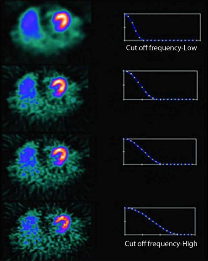
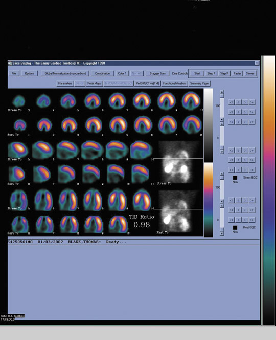
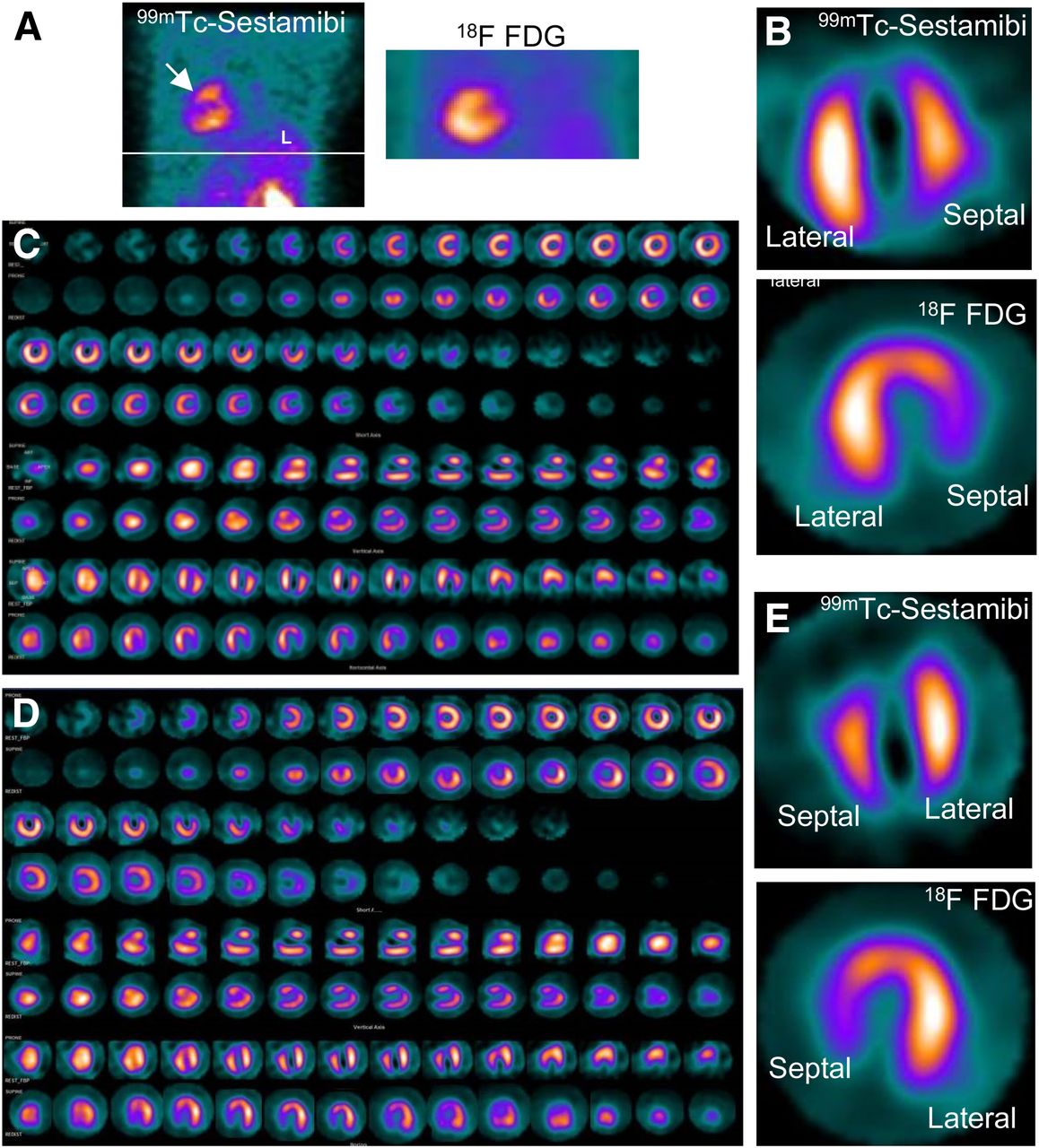
Imaging of dextrocardia in humans requires an understanding of the orientation of the heart chambers and walls. There are many types of cardiac malpositioning, such as dextrocardia (with or without situs inversus), mesocardia, and levocardia. Myocardial perfusion scintigraphy of dextrocardia has been explained in case reports and imaging atlases; however, myocardial viability assessment using nuclear medicine imaging techniques is less documented in the literature. Methods: In 2 cases of dextrocardia with situs inversus and 1 case of mesocardia, myocardial viability was assessed using 99mTc-sestamibi rest perfusion scintigraphy and 18F-FDG PET. Cardiac SPECT images of dextrocardia with situs inversus were acquired using the feet-first supine position with a 180° arc from left anterior oblique to right posterior oblique, whereas a right-lateral–to–left-lateral arc was used for mesocardia. The processing and reconstruction were done by entering the dataset for the feet-first supine position and repeating after entering the dataset for the feet-first prone position. The 2 sets of reconstructed images were compared for orientation of walls and cardiac chambers. Results: The first processing, using the feet-first supine position, revealed an interchanged septum and lateral wall in reconstructed images of dextrocardia with situs inversus. This interchange was corrected by changing the position to prone during processing of the rest perfusion and PET raw data. The display of cardiac slices in various axes matched the conventional nomenclature for the septum and lateral wall, leading to easy interpretation. However, this change was not required in the mesocardia, for which the location of the heart chambers was not interchanged. Conclusion: Because the acquisition protocol for SPECT is a semicircular orbit, the various types of dextrocardia require careful selection of the arc, with the patient positioning kept feet-first supine. Processing and reconstruction of data by changing the patient position to prone was found to be most useful method of matching the septum and lateral wall orientation for interpretation of images.
Single Photon Emission Computed Tomography (SPECT) Myocardial

Myocardial perfusion single photon computed tomography: An Atlas

Approach to Dextrocardia in Adults: Review
Myocardial viability • APPLIED RADIOLOGY

Tl-201 treadmill stress/rest myocardial perfusion SPECT short axis
Assessment of Myocardial Viability Using Nuclear Medicine Imaging
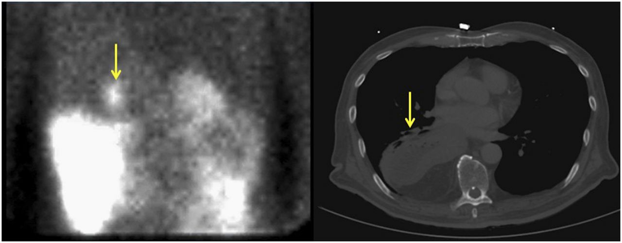
Incidental Findings on Myocardial Perfusion SPECT Images

The Impact of the Coronavirus Disease 2019 Pandemic on the

Approach to Dextrocardia in Adults: Review
de
por adulto (o preço varia de acordo com o tamanho do grupo)